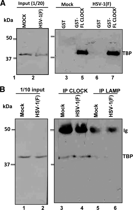
Fig. 1.
The TBP subunit of TFIID exists in complex with CLOCK. (A) Lysates from mock- or HSV-1(F)-infected (10 PFU/cell) HEp-2 cells (2 × 10⁷), harvested at 8 h after infection, were mixed with an equal amount of GST or GST-CLOCK purified proteins immobilized on glutathione beads. The bead-bound complexes were electrophoretically separated on 9% polyacrylamide gels, transferred to nitrocellulose sheets, and immunoblotted with the TFIID (TBP) mouse monoclonal antibody, as described in Materials and Methods. (B) HEp-2 cells (5 × 10⁶) were mock infected or exposed to 10 PFU of HSV-1(F) per cell. The cells were harvested 8 h after infection and reacted with CLOCK or LAMP-1 rabbit polyclonal antibodies. The electrophoretically separated immune precipitates were probed with anti-TFIID (TBP) antibody as described above.

