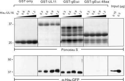
Fig. 3.
Direct-interaction assays with UL16 and gEtail. The indicated amounts of purified, soluble His6-UL16 protein were mixed with the indicated GST fusion proteins immobilized on glutathione beads and incubated to allow binding. The beads were washed to remove unbound His6-UL16, and the samples were subjected to SDS-PAGE. Two samples of input His6-UL16 were included to show the efficiency of binding (rightmost lanes). The separated proteins were transferred to a nitrocellulose membrane, and the GST-fusion proteins were visualized by Ponceau S staining (upper panel). Bound and input His6-UL16 proteins were detected by immunoblotting with an antibody that recognizes the His6 tag (bottom panel).

