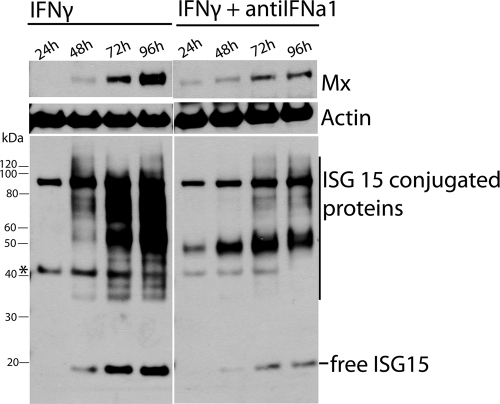
Fig. 9.
Time course study of Mx and ISG15 protein expression was conducted in TO cells stimulated with 100 ng of IFN-γ/ml with or without 1% antiIFNa1 anti-serum. Lysates were harvested at the indicated time points ranging from 24 to 96 h and subjected to Western blot analysis. The blot was incubated with antibodies against Mx (1:3,000) and actin (1:1,000) and developed before stripping and reincubation with antibody against ISG15 (1:20,000). The asterisk indicates the remnants of actin antibody, which was not completely removed during the stripping procedure.

