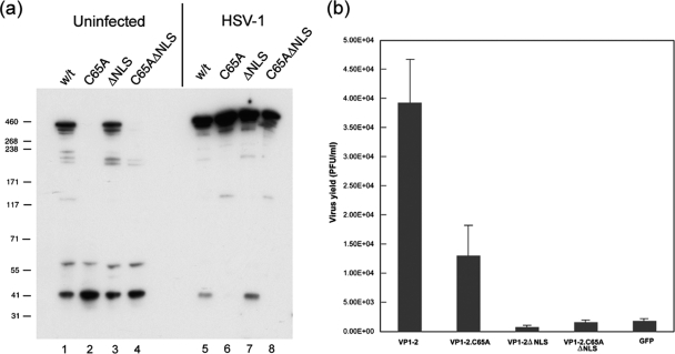
Fig. 7.
Relative instability of full-length VP1-2.C65A is rescued by replication-defective virus. (a and b) Cells were transiently transfected with expression vectors for full-length VP1-2 or mutant versions as indicated. Duplicate sets were either mock infected or superinfected with the VP1-2-negative deletion mutant, KΔUL36 (MOI 5), and further incubated for 16 h, after which time cells were harvested and processed for expression levels of VP1-2 (a) or recovery of virus growth by plaque titration in HS30 cells (b). Virus infection resulted in a significant increase in all species of VP1-2 but in particular altered the ratio of the C65A mutants, whose levels were now overall similar to the wt VP1-2, unlike what was seen for uninfected cells. As described previously, VP1-2 substantially complemented growth of KΔUL36. Background levels seen with the control vector expressing green fluorescent protein (GFP) are due to the low level of revertant virus obtained in KΔUL36 preparation (11). VP1-2.ΔNLS showed no complementation above background level. However, VP1-2.C65A exhibited significant levels of complementation above background and only 2- to 3-fold lower than that for wt VP1-2.

