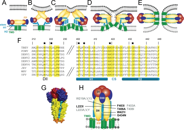
Fig. 1.
Representations of the flavivirus fusion model (A to E) and sequence alignment of the stem regions of the E fusion proteins of different flaviviruses (F) and the postfusion E trimer (G and H). Color coding for E is as follows: red, domain I (DI); yellow, domain II (DII); blue, domain III (DIII); orange, fusion peptide; cyan, stem helix (helix 1 [H1] and H2); green, transmembrane domain (TMD). Color coding for membranes is as follows: light gray, virus; dark gray, cell. (A) E dimer on the virion surface. (B) Low-pH-induced E dimer dissociation, lifting of the stem, and FP interaction with the target membrane. (C) DIII relocation to the side of the molecule, E protein trimerization, and initiation of stem zippering. (D) Hemifusion intermediate with fused outer leaflets. (E) Postfusion hairpin-like E trimer with FPs and TMDs at the same side of the molecule. (F) Amino acid sequence alignment of DIIs (amino acids 212 to 230) and stem regions (amino acids 402 to 449) of several flavivirus E proteins (TBEV numbering): TBEV (GenBank accession number U27495), Powassan virus (POWV) (GenBank accession number L06436), dengue virus (DENV) type 1 (GenBank accession number FJ687432), DENV type 2 (GenBank accession number NC_001474), DENV type 3 (GenBank accession number FJ850055), DENV type 4 (GenBank accession number AY618990), Japanese encephalitis virus (GenBank accession number EF571853), West Nile virus (WNV) (GenBank accession number DQ211652), and yellow fever virus (YFV) (GenBank accession number AY640589). Identical amino acids are highlighted in yellow. Residues which were mutated in this study are labeled with black stars. The predicted stem helices and the conserved sequence (CS) between the helices are indicated below the alignment. (G) Surface representation of truncated sE postfusion trimer of TBEV (side view) (Protein Data Bank [PDB] accession number 1URZ). Stem helices added schematically to the structure are shown as cylinders in a groove provided by neighboring DII domains. (H) Schematic representation of the full-length E trimer (side view), displaying the mutations introduced into the stem (right side) and DII (left side). Mutations that gave rise to mature particles are labeled in black, and mutations that abolished either particle secretion or maturation are labeled in gray.

