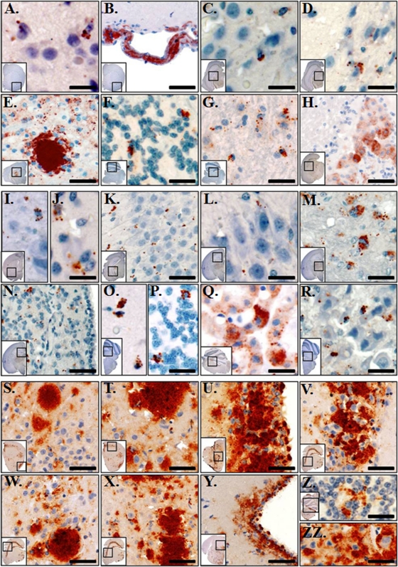
Fig. 2.
Prairie voles accumulate PrPRES in diverse brain regions. (A to H) Prairie voles inoculated with CWD-infected deer brain D10. In the forebrain, PrPRES (red staining) was heaviest in the ventral and medial gray matter of the cerebral cortex and was multifocal to regionally extensive (A). Clumped aggregates of PrPRES were seen in the tunica adventitia of many, mostly meningeal, vascular channels in the forebrain of 2/3 IHC-positive (IHC+) animals (B). Scant to minimal PrPRES was seen in the hippocampi of 3/3 IHC+ animals, with fine to coarsely granular aggregates in the CA1-CA3 layers (C) and in the stratum granulosum (D). Coarsely granular to clumped, consistently cell-associated aggregates of PrPRES are shown in the thalamus and hypothalamus (E). In the cerebellum, PrPRES was identified in 3/3 IHC+ animals and was consistently cell associated and clumped within the granular layer (F). In the brain stem, multifocal to regionally extensive accumulations of PrPRES were identified in 3/3 IHC+ animals, which was both cell and neuropil associated (G). In 1/3 IHC+ animals, heavy amounts of granular to clumped PrPRES was identified in the ependymal epithelial cells (H). (I to R) Prairie voles inoculated with vPMCA-PrPRES. In the forebrain, PrPRES was identified in 8/8 IHC+ animals and was heaviest in the gray matter of the cerebral cortex including the ventral and medial gray matter (I). In 4/8 IHC+ animals, scattered mild to moderate, fine to coarsely granular PrPRES was identified within the putamen and caudate nucleus (J). In the hippocampus, scant to minimal PrPRES was seen in 5/8 IHC+ animals with fine to coarsely granular aggregates seen in the CA1-CA3 layers (K) and in the stratum granulosum (L). In the thalamus and hypothalamus, coarsely granular to clumped, consistently cell-associated aggregates of PrPRES were identified in 8/8 IHC+ animals (M). In 4/8 IHC+ animals, coarsely granular to clumped aggregates of PrPRES were identified in the medial habenular nucleus (N). In 8/8 IHC+ animals, cell-associated and clumped PrPRES was consistently identified in the granular layer of the cerebellum (O). In 2/8 animals, sparse depositions of PrPRES were seen in the molecular and Purkinje layers (P). In 3/8 IHC+ animals, heavy amounts of granular to clumped PrPRES were identified in the ependymal epithelial cells (Q). In the brain stem, multifocal to regionally extensive accumulations of granular to clumped PrPRES were identified in 8/8 IHC+ animals, which was both cell associated and regionally disseminated throughout the neuropil (R). (S to ZZ) Tg(CerPrP)1536 mice inoculated with CWD-infected deer brain D10. Similar patterns of PrPRES immunoreactivity were observed in 7/7 evaluated animals. In the forebrain, PrPRES was extensively distributed throughout the gray matter of the cerebral cortex (S and T). Heavy amounts of clumped PrPRES were seen in the caudoputamen, with the densest aggregates within the periventricular gray matter and ependymal epithelial cells (U). Heavy, clumped, and largely extracellular PrPRES aggregates were observed throughout the entirety of the hippocampal formation, including the pyramidal layer (V), the stratum radiatum of field CA1 (W), and field CA2 (X). Moderate to marked, finely granular to clumped PrPRES deposits were seen in the periaqueductal gray matter (Y). Cell and non-cell-associated PrPRES was observed in the granular (Z) and molecular (ZZ) layers of the cerebellum. Scale bars, 25 μm (A, C, D, I, J, L, and R), 40 μm (F, G, K, M, O, P, Q, Z, and ZZ), 60 μm (E, N, S, T, U, V, W, X, and Y), and 75 μm (B and H).

