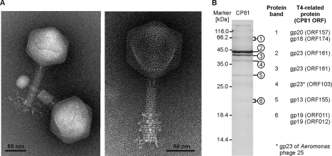
Fig. 1.
Morphology and structural proteins of CP81. (A) Electron micrographs of negatively stained virions. The micrographs on the left and on the right show particles stained with phosphotungstate and ammonium molybdate, respectively (see Materials and Methods). The head diameter was measured between the sides parallel and diagonal to the tail. Both axes were identical within the error range. A collar structure as in T4 was not observed. (B) SDS-PAGE of structural CP81 proteins (27). As a marker, the unstained molecular mass marker of Fermentas (St. Leon-Rot, Germany) was used. Visible bands were excised from the gel and analyzed by mass spectrometry (53).

