Fig. 3.
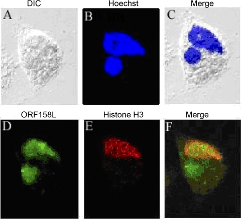
Colocalization of ORF158 with histone H3. (A) A cell in a DIC image. (B) DNA staining with Hoechst (blue) in the nucleus and the virus assembly center (round shape). (C) Merged image of DIC and Hoechst staining. (D) ORF158L is labeled with Alexa Fluor 488 (green). (E) Histone H3 is labeled with Alexa Fluor 594 (red). (F) Colocalization of ORF158L and histone H3 (yellow). ORF158L is accumulated in the host nucleus and viral assembly center (green area in the cytoplasm). Histone H3 and ORF158L proteins are colocalized in the nucleus.

