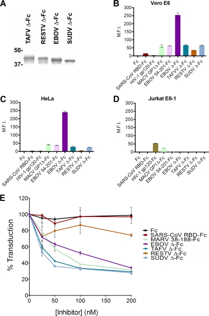
Fig. 2.
Expression, cell surface binding, and cell entry-inhibitory effect of TAFV, SUDV, RESTV, and EBOV Δ-peptide-Fcs. (A) EBOV, RESTV, SUDV, and TAFV Δ-Fc were purified from transfected cells and visualized by Coomassie staining. (B, C, and D) Δ-Peptide-Fcs or control proteins (100 nM) were incubated with Vero E6 or HeLa cells or with Jurkat E6-1 lymphocytes and analyzed by flow cytometry. (E) The indicated concentrations of Δ-Fcs or control proteins Fc, SARS-CoV RBD-Fc, or MARV 38-188-Fc were incubated with Vero E6 cells and eGFP-expressing MLV pseudotyped with MARV GP1,2. Entry of pseudotyped MLV was quantified by measuring green fluorescence. Error bars indicate standard deviations.

