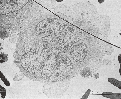
Figure 2.
Transmission electron micrograph of a human megakaryocyte in Stage I. The specimen was obtained by marrow aspiration. A MK, probably in endomitosis stage, has a multilobulated large nucleus and abundant free ribosomes, mitochondria of large size with electron translucent matrix, and prominent Golgi body. Upper left side: a part of a MK with platelet specific granules, right side: a circulating platelet attached to the MK is seen. (Reproduced from Kosaki and Fujimoto6) Fig. 1.50 with permission from the publisher)

