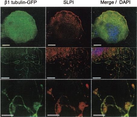
Figure 11.
Immunofluorescence (IF) analysis of primary MKs expressing a β1-tubulin-GFP fusion protein, SLPI (secretory leukocyte protease inhibitor) is expressed naturally in MKs. Cellular SLPI is detected by a specific antibody and Texas red-labelled secondary antibody. First column; IF analysis, second column; immunostaining of SLPI, third column; merge, including nuclear DAPI staining. Each row represents different cells. Scale bars, 10 µm (Reproduced from Schulze et al.60) Fig. 1B, with permission from the publisher)

