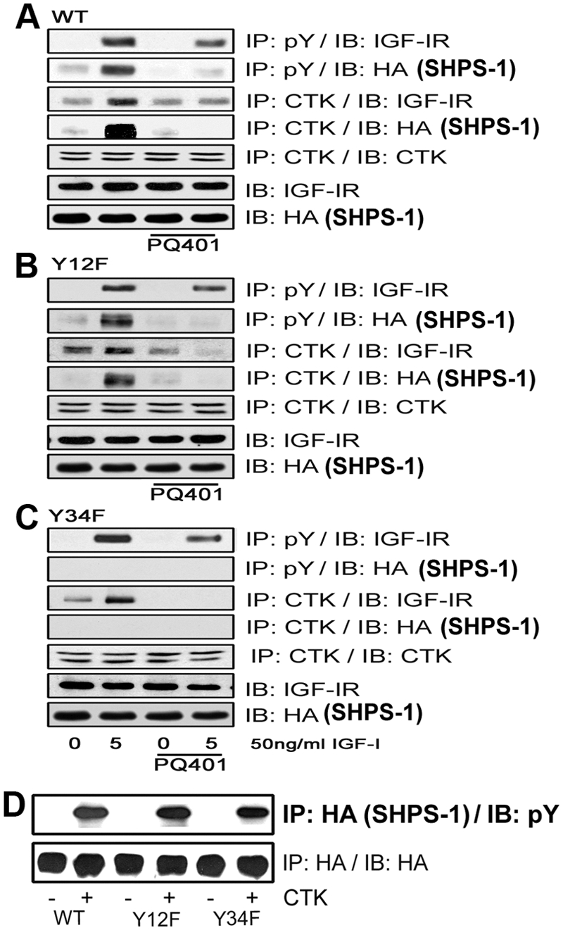
Fig. 4.

IGF-IR and CTK phosphorylate specific tyrosines on SHPS-1. A–C, Quiescent VSMC expressing SHPS1-WT (A), Y12F (B), and Y34F (C) were cultured and maintained in DMEM-HG and then serum starved overnight before being incubated with or without the IGF-IR tyrosine kinase inhibitor, PQ401 (10 μm), for 1–2 h. IGF-I was added for 5 min. Cell lysates were immunoprecipitated (IP) with an anti-pY antibody and then immunoblotted (IB) for IGF-IR (first panel) or HA (second panel). Similarly, cell lysates from the same experiments were immunoprecipitated with an anti-CTK antibody and then immunoblotted with anti-IGF-IR (third panel), anti-HA (fourth panel), or anti-CTK antibodies (fifth panel). Twenty micrograms of protein from the same whole-cell lysate were directly immunoblotted with an anti-IGF-IR antibody (sixth panel) or with an anti-HA antibody (seventh panel). D, Confluent SHPS-WT, Y12F, and Y34F cells were serum starved for 16 h in DMEM-HG, and the cell lysates were immunoprecipitated with anti-HA antiserum. The immunoprecipitates were incubated with or without CTK to determine HA-tagged SHPS-1 phosphorylation in vitro. After centrifugation the resultant supernatants were immunoblotted using a pY antibody (upper panel) or an HA antibody as a loading control (lower panel).
