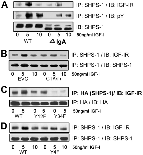
Fig. 7.
IGF-IR/SHPS-1 association and IGF-IR-stimulated SHPS-1 phosphorylation is mediated in part by CTK. A, Confluent VSMC expressing SHPS-1WT or SHPS-1 IgA deletion mutant were serum starved for 16 h in DMEM-HG and then exposed to IGF-I for the times indicated. Cell lysates were immunoprecipitated (IP) with an anti-SHPS-1 antibody and then immunoblotted (IB) for IGF-IR (top panel) or with an anti-pY antibody (middle panel). Twenty micrograms of protein from the same or similar whole-cell lysate were directly IB with an anti-SHPS-1 antibody (lower panel). B, Confluent empty control vector (EVC), and CTK shRNA cell cultures were serum starved for 16 h and IGF-I (50 ng/ml) was added for times indicated. The cell lysates were immunoprecipitated with an anti-SHPS-1 antibody and then immunoblotted for IGF-IR or using an anti-SHPS-1 antibody. C, Confluent VSMC-expressing SHPS-1-WT and SHPS-1 (Y12F and Y34F) mutant cells were serum starved for 16 h in DMEM-HG and then exposed to IGF-I for 5 min. The extent of HA-tagged SHPS-1 association with IGF-IR was determined by immunoprecipitating HA and immunoblotting for IGF-IR or with an anti-HA antibody as a loading control. D, Confluent VSMC-expressing IGF-IR-WT and IGF-IR-Y4F mutant cells were serum starved for 16 h in DMEM-HG and then exposed to IGF-I for the indicated period of time. Cell lysates were immunoprecipitated with an anti-SHPS-1 antibody and immunoblotted using an anti-IGF-IR antibody or an anti-SHPS-1 antibody.

