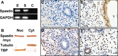
Fig. 1.
Spastin is expressed in the adult human reproductive tract. A, Total RNA was obtained from Ishikawa cells as well as primary HESC and expression of spastin was evaluated by RT-PCR and normalized to glyceraldehyde-3-phosphate dehydrogenase (GAPDH). Spastin was expressed in both Ishikawa endometrial epithelial cells (E) as well as HESC (S). Lane C represents the negative control (water). B, Spastin protein expression was evaluated in Ishikawa endometrial epithelial cells transfected with pMT21/spastin-myc by Western blot analysis. For Western blot analysis, nuclear as well as cytoplasmic extracts were derived from Ishikawa cells and evaluated separately using antispastin. Spastin (65 kDa) was expressed in both Ishikawa nuclear (Nuc) as well as cytoplasmic extracts (Cyt). Anti-TBP and anti-α-tubulin were used as nuclear and cytoplasmic-compartment-specific markers. C–F, Endogenous uterine spastin expression was evaluated in intact human endometrium using antispastin, by immunohistochemistry. Spastin expression was evaluated on d 12 (D), 17 (E), and 24 (F) of the human menstrual cycle. C, Negative control using mouse IgG. D–F, Spastin was expressed in both the nucleus and cytoplasm of endometrial epithelial cells as well as stromal cells (D–F). Stromal spastin remained unchanged from the proliferative phase of the menstrual cycle (D) to the secretory phase (E and F). In contrast, epithelial spastin expression was observed only in the progesterone driven secretory phase of the menstrual cycle (E and F). All magnifications, ×100 (C–F).

