
Figure 8.
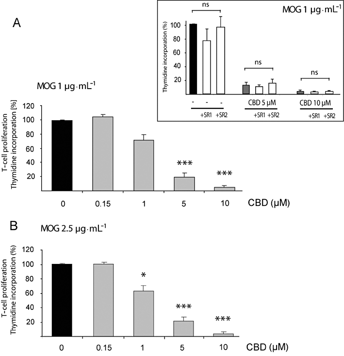
Cannabidiol (CBD) inhibits MOG-induced T-cell proliferation. MOG-induced T-cell proliferation was determined by [3H]thymidine incorporation. Encephalitogenic T cells were co-cultured with antigen presenting cells and exposed to MOG35-55 at 1 (A) and 2.5 µg·mL−1 (B). CBD was added 5 min before MOG. Assays were carried out each time in triplicate and mean per cent values ± SEM are shown based on four independent experiments. (A) One-way anovaF(4,19) = 124.7, P < 0.0001; (B) One-way anovaF(4,19) = 31.1, P < 0.0001, followed by Bonferroni post hoc test. *P < 0.05, ***P < 0.001 versus MOG alone at the respective concentration. Inset: neither the CB1 receptor antagonist (1 µM of SR141716; SR1) nor the CB2 receptor antagonist (1 µM of SR144528; SR2) applied 30 min before CBD and MOG affected MOG35-55 (1 µg·mL−1) – induced T-cell proliferation either in the absence or presence of CBD; one-way anovaF(8,26) = 26.8, P < 0.05; ns, non-significant.