
Figure 2.
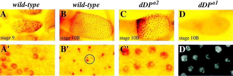
dDP mutants affect amplification and the shutoff of genomic replication. (A–D) DNA replication was assayed by BrdU labeling observed by antibody staining detected by the HRP reaction. BrdU incorporation is indicated by the red staining. (A–D) The same magnification, and shown below (A′–D′) at a higher magnification. (A,A′) In stage 9 control dDPa1/+ egg chambers genomic replication is asynchronous among follicle cell nuclei. The egg chamber contains follicle cells that are labeled (S phase) with BrdU and those that are not labeled (G phase). The same pattern of nuclear BrdU incorporation is observed in the female-sterile dE2F and dDP mutants. (B,B′) In stage-10B control egg chambers, polyploidization of the follicle cell genome is no longer observed. Instead, synchronous subnuclear BrdU incorporation is seen. The subnuclear dots correspond to amplifying chorion clusters. The nucleus of a single follicle cell is indicated by a circle. The different intensity and size of foci (one large, one medium, and two small) reflect the extent of gene amplification (Calvi et al. 1998). (C,C′) A stage-10B dDPa2/Df; hs–dDP/+ mutant egg chamber is shown. Genomic replication is occurring synchronously in the follicle cells of this mutant egg chamber. The same phenotypes were observed for the dDPa1/Df mutant. Note that the nuclei are much larger than at stage 9, indicating higher ploidy due to inappropriate genomic replication. (D) A stage-10B dDPa1/Df egg chamber is shown. Genomic replication has been shutoff, but amplification is not observed. The same replication defect was observed for dDPa2/Df; hs–dDP/+. (D′) A magnified DAPI image of D showing that the follicle cell nuclei are present and polyploid.