
Figure 6.
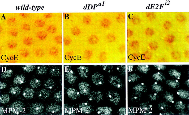
Cyclin E levels and activity are not affected in the dDP and dE2F mutants. A field of follicle cells from a stage-10B egg chamber is shown in each panel. (A–C) Cyclin E protein levels in wild-type and mutant follicle cells following staining with a monoclonal antibody (Richardson et al. 1995). The antibody staining is detected by the HRP reaction. (A) In wild-type follicle cells cyclin E is present at uniformly high levels in all the nuclei. (B,C) The levels of Cyclin E protein appear normal in mutant dDPa1/Df and dE2Fi2/Df follicle cells. (D–F) MPM2 staining reflects cyclin E activity in follicle cells (Calvi et al. 1998). MPM2 staining is visualized by confocal immunofluorescence. (D) In wild-type follicle cells a bright nuclear sphere is seen in all the follicle cell nuclei. (E,F) An identical MPM2 sphere is observed in the mutant dDPa1/Df and dE2Fi2/Df follicle cells.