
Figure 8.
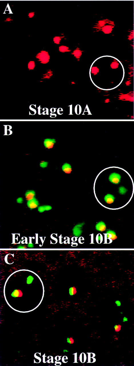
ORC2 and BrdU double labeling. Egg chambers were double labeled with anti-ORC2 and BrdU and staged according to Spradling (1993). ORC2 staining is shown in red and BrdU labeling in green. A field of follicle cells is shown in each panel. (A) In stage-10A egg chambers two large foci of ORC2 staining are present in each follicle cell nucleus (designated by a circle), but there is no BrdU incorporation. (B) Early stage-10B egg chambers are small, only slightly larger than stage 10A, and in these the two large ORC2 foci coincide with sites of BrdU incorporation. (C) In stage-10B egg chambers that are large and developmentally later, only a single focus of ORC2 is present and it corresponds to the largest dot of incorporated BrdU.