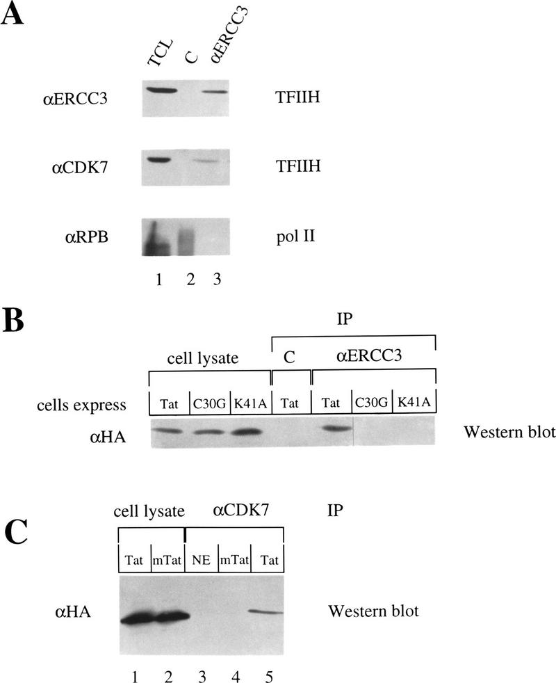
Figure 1.
Tat associates with TFIIH in the absence of the Pol II holoenzyme in vivo. (A) Total cell lysates (TCL) from COS cells were immunoprecipitated with αCIITA (lane 2) or αERCC3 antibodies (lane 3). One-third of the lysate was used as the input control (lane 1). Samples were separated by SDS-PAGE, transferred to membranes, and probed with the antibodies indicated on the left. (B) COS cells expressed HA-tagged wild-type (Tat) or mutant (C30G or K41A) Tat proteins. Cell lysates were immunoprecipitated with either the αCIITA (C) antibody or the αERCC3 antibody (αERCC3). Equal amounts of the lysates were loaded as input controls. Samples were processed as in A and probed with the αHA antibody. (C) Nuclear extracts (NE) or lysates from cells expressing HA-tagged wild-type Tat (Tat) or mutant Tat (mTat = K41A) were immunoprecipitated with the αCDK7 antibody. One-half of these cell lysates were loaded as input controls (lanes 1,2). Samples were processed and blots probed with αHA antibodies as described above.

