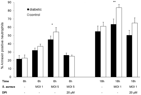
Figure 5. Neutrophil apoptosis—ex vivo infection.
Apoptosis of neutrophils isolated from mouse peritoneal cavities after injection of casein as a sterile inflammatory stimulus. The cells were incubated ex vivo for 6 or 18 h with or without the addition of opsonized staphylococci. In the absence of S. aureus, no significant differences in annexin binding were observed. However, at a S. aureus MOI of 1, significantly fewer neutrophils from diabetic mice were apoptotic after 18 h; an MOI of 5 resulted in significant differences between the mouse groups by 6 h. No significant differences between neutrophils from diabetic and nondiabetic mice were found (at 6 h or 18 h) in the presence of 20 µM DPI. Mean percent apoptotic neutrophils ± SEM from 3 to 6 independent experiments are depicted. P values are calculated from pair-wise comparisons of age-matched mice, * indicates P<0.05, ** P<0.01.

