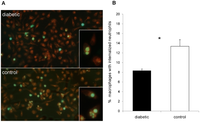
Figure 7. In vitro phagocytosis of apoptotic neutrophils by murine macrophages.
Inflammatory cells were isolated from infected mouse peritoneal cavities 18 h after IP challenge with 108 CFU PS80. The neutrophils were labeled with CellTracker Green, incubated in vitro for 8 h to induce apoptosis, and incubated with adherent autologous macrophages for 90 min to allow for uptake. (A) Representative image of CellMask Orange-stained macrophages with phagocytosed neutrophils from an age-matched pair of a diabetic (upper picture) and nondiabetic (lower picture) control mice. Phagocytosis of apoptotic neutrophils by macrophages from diabetic mice is less than that of macrophages from nondiabetic mice. Some neutrophils are still extracellular whereas others have been internalized. (B) Macrophages from diabetic mice ingested significantly fewer neutrophils than macrophages from nondiabetic control mice. Data are means ± SEM of six independent experiments; * indicates P<0.05.

