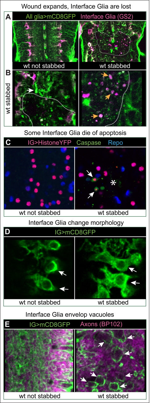
Figure 2. Glial responses to injury.
(A) Injury affects most prominently the Interface Glia (magenta), visualised with GS2. Longitudinal views of the VNC, dashed lines delineate wound edges. (B) Higher magnification views of (A) showing loss of GS2+ glia upon injury, accumulation of glial material (white arrow), and GS2+ glial debris (orange arrows). (C) Some Interface Glia die of apoptosis, as the cytoplasmic apoptotic marker cleaved-Caspase-3 surrounds Interface Glial nuclei labelled with Repo and Histone-YFP expressed in IG only with alrmGAL4 (arrows). Asterisk indicates lesion site. (D) Glia project more filopodia and lamellipodia (arrows) after injury compared to non-stabbed controls. Glial membranes are visualised with mCD8GFP and anti-GFP. (E) Interface Glial processes envelop vacuoles that form in the axonal neuropile after injury (arrows). All axons are labelled with BP102 antibodies. Anterior is up. Genotypes: (A,B) all glia: w; UASmCD8GFP/+;repoGAL4/+. (C,D,E) Interface Glia: (C) IG nuclei: w;alrmGAL4/UAShistoneYFP; (D,E) glial processes and membranes w;alrmGAL4/UASmcD8GFP.

