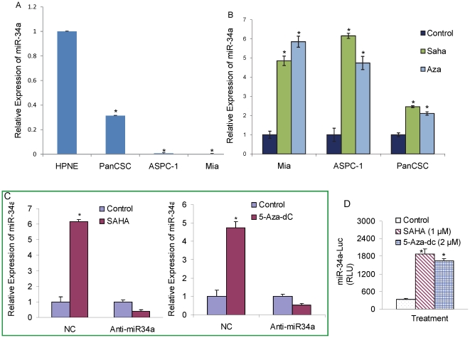
Figure 1. Restoration of the expression of miR-34a in pancreatic cancer cells.
(A) Relative expression of miR-34a was quantified in human pancreatic cancer cell lines ASPC-1(p53wt), MiaPACA-2 (p53mutant), human pancreatic cancer stem cells (PanCSC) and human pancreatic normal ductal epithelial cells (HPNE). (B) Restoration of miR34a by SAHA and Aza-5dC. Pancreatic cancer cell lines ASPC-1(p53wt), MiaPACA-2 (p53mutant) and pancreatic cancer stem cells were treated with SAHA (3 µM) or Aza-5dC (4 µM) for 24 h. The expression of miR-34a was quantified using quantitative reverse transcriptase polymerase chain reaction (RT-PCR-Taqman) and Taqman Real Time Assays, and normalized to RNU48 expression. Data represent mean ± SD. * = significantly different from control, P<0.05. (C), Anti-miR34a inhibits the ability of SAHA and Aza-5dC to restore miR34a. Pancreatic CSCs were transiently transfected with either negative control (scrambled) or anti-miR34a oligonucleotide, and treated with SAHA (3 µM) or Aza-5dC (4 µM) for 24 h. RNA was extracted to measure the expression of miR34a by q-RT-PCR as described above. NC = negative control. (D), miR34a-Luciferase reporter activity. Pancreatic CSCs were transfected with either negative control (scrambled) or anti-miR34a oligonucleotides along with miR34a-Luc construct, and treated with either SAHA (1 µM) or Aza-5dC (2 µM) for 24 h. Luciferase activity was measured as per manufacturer's instructions (Promega). Data represent mean ± SD. * = significantly different from control, P<0.05.

