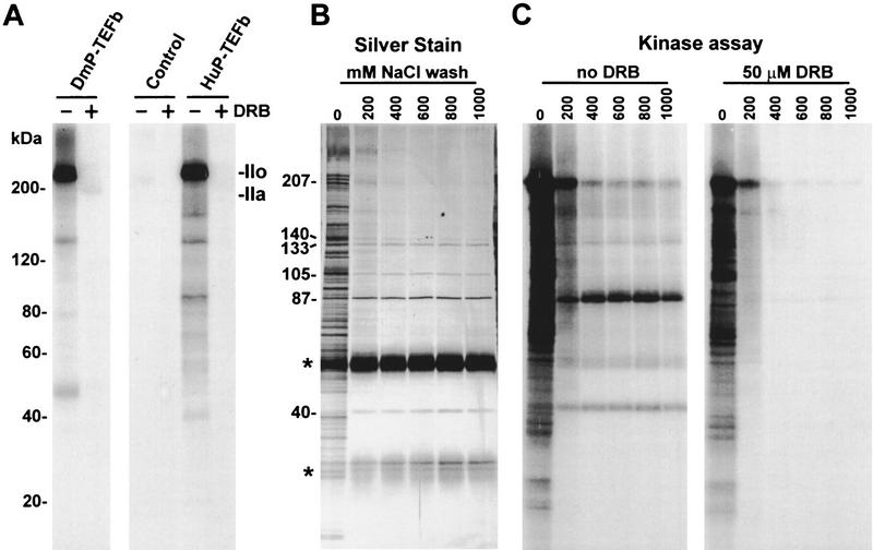
Figure 3.
Immunoprecipitation of human P-TEFb. (A) CTD kinase assay. Purified Drosophila RNA polymerase II (Price et al. 1987; Marshall and Price 1995) was used as substrate. (DmP-TEFb) Drosophila P-TEFb; (Control) beads after depletion of HNE by affinity-purified rabbit anti-goat IgG; (HuP-TEFb) beads after depletion of HNE by PITALRE–CT IgG. Products were analyzed by 6%–15% gradient SDS-PAGE. Selected size standards from the 10-kD ladder are indicated and apply to all gels. (B) Silver-stained SDS-PAGE of proteins bound to PITALRE–CT IgG beads after washing with a buffer containing 20 mm HEPES (pH 7.6), 0.5% NP-40, 1% Triton X-100, and 5 mm DTT and the indicated amount of NaCl. Sizes of proteins remaining after high salt wash are indicated. Bands marked with an asterisk (*) are from IgG. (C) Kinase assay with only endogenous substrates of the fractions analyzed in B either without or with 50 μm DRB, as indicated.

