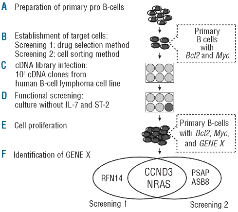
Figure 3.
Diagram of the retroviral cDNA expression library screening protocol. (A) Mouse primary pro B-cells were purified from fetal liver of BALB/c mice. (B) Bcl2 and Myc-expressing target cells were established by the drug selection method for Screening 1 and the cell sorting method for Screening 2. (C) Target cells were infected with a retroviral expression library containing 106 cDNA from SU-DHL-6. (D) Functional screenings were performed under culture condition in the absence of IL-7 and ST-2 cells. (E) Proliferation of cells transduced with GENE X, which cooperates with Bcl2 and Myc. (F) Inserted cDNA were recovered by PCR-amplification using the retro-viral-specific primers from retrovirus-integrated genomic DNA of proliferating cells. CCND3 and NRAS were identified in both screenings.

