
Figure 2.
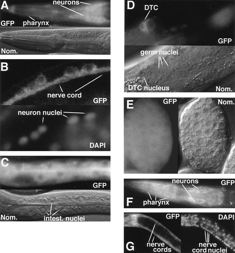
DAF-3 and DAF-4 expression. All DAF-3 photographs show animals with the full-length functional DAF-3/GFP fusion gene shown in Fig. 3. In A and C–F, intrinsic fluorescence is used to visualize GFP; in B and G, anti-GFP antibodies are used. (A) DAF-3/GFP head expression in an L1 animal. Weak DAF-3/GFP expression in the pharynx impedes cell identification, but the main body of the pharynx is filled, implying expression in pharyngeal muscle. (B) DAF-3/GFP expression in the ventral nerve cord of an adult animal. L1 animals look similar. (C) DAF-3/GFP expression in the intestine of an L1 animal. (D) DAF-3/GFP expression in the distal tip cell of an L4 animal. (E) DAF-3/GFP expression in an embryo with ∼200 nuclei. (F) DAF-4/GFP expression in the head of an L1 animal. (G) DAF-4/GFP expression in the dorsal nerve cord and ventral nerve cord of an L4 animal. Although the intestine can be seen in this picture, the DAF-4/GFP is not visible, because in this focal plane, the intestinal cell membrane is obscured by the bright fluorescence in the nerve cords.