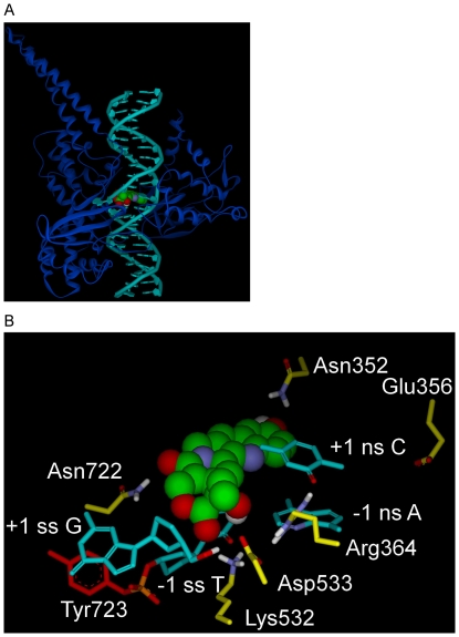
Figure 2. Rotated +1 Nucleoside model for Top1 interaction with dsDNA and 10-OH CPT.
A) Rotated +1 Nucleoside model, Top1 shown as blue ribbon, dsDNA in teal with the rotated +1 deoxyguanosine left of 10-OH CPT (CPK rendering; carbon, green; oxygen, red; nitrogen, blue; hydrogen, white). B) Rotated +1 Nucleoside model, close up of Fig. 2A active-site. 10-OH CPT with E-ring in foreground, bound in the Top1/dsDNA active-site in which the +1 scissile strand G is rotated out of the helix to the left until trapped in a network of H-bonds/electrostatic interactions with Asp533 (center) and Arg488/Arg590 (not shown for clarity, see Fig. 4A). Selected atoms involved in H-bonds/electrostatic interactions are colored: nitrogen, blue; oxygen, red; hydrogen, white. Top1 side-chain carbons are yellow, except for Tyr723 (red) in tyrosyl-phosphate bond (phosphorus, orange) to the -1 scissile strand T. 10-OH CPT interactions: 10-OH CPT D-ring stacks over the -1 scissile strand T; 20-OH H-bonds to -1 scissile strand T carbonyl oxygen; A-ring 10-OH oxygen makes electrostatic interaction with Asn352 nitrogen (3.6 Å); E-ring carbonyl oxygen H-bonds to Lys532 nitrogen; C-ring carbonyl oxygen H-bonds to Asn722 nitrogen. Scissile strand rotated +1 G 5′OH H-bonds to Asp533. Arg364 nitrogens H-bond to +1 non-scissile strand C carbonyl oxygen and -1 non-scissile strand A nitrogen. Scissile strand, ss; non-scissile strand, ns. For flat image see Fig. 4A.

