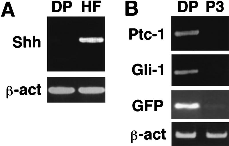
Figure 2.
DP cells respond to Shh. (A) After dissociation of newborn skin, RT–PCR revealed Shh mRNA in the GFP-negative population (HF) that includes the follicular epithelium. Shh mRNA was not detected in the GFP-positive population (DP). β-actin (β-act) serves to compare input RNA levels in this and subsequent panels. (B) ptc and Gli1 are expressed in freshly isolated DP cells, but transcript levels decrease upon passage in culture (P3), as do those from the GFP transgene.

