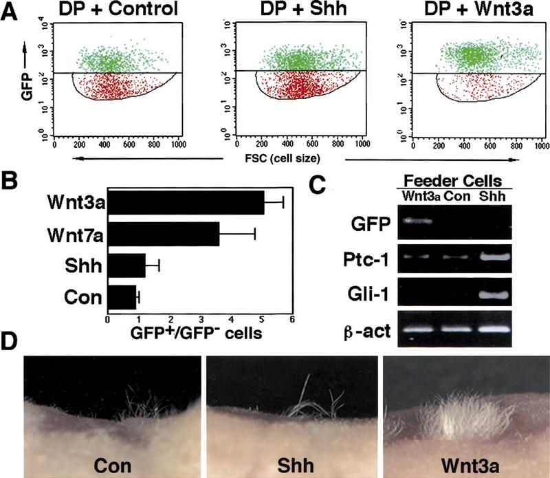
Figure 3.

Wnt signaling to DP cells maintains hair inductive activity. (A) Flow cytometric analysis of DP cells cocultured with feeder cells producing Wnt3a or Shh, or infected with a control vector. The horizontal line demarcates the division between GFP-positive (green) and -negative (red) DP cells. The feeder cells found below the lower gate were excluded from this analysis. (B) The ratio of GFP-positive to GFP-negative cells is shown for each treatment. (C) RT–PCR analysis of cocultured DP cells. GFP mRNA levels were maintained by exposure to Wnt3a but not Shh or control feeder layers. ptc and Gli1 expression was induced in DP cells cocultured with Shh-expressing feeder cells. DP cells cultured with control feeders that do not express Shh or feeders expressing Wnt3a express low levels of ptc and undetectable levels of Gli1. (D) Wnt3a-treated cells maintain hair inductive activity. Three weeks after grafting to nude mouse hosts, only occasional hairs were observed in grafts of control or Shh-treated DP cells. DP cells exposed to Wnt3a directed formation of a dense patch of hair in the graft.
