
Figure 3.
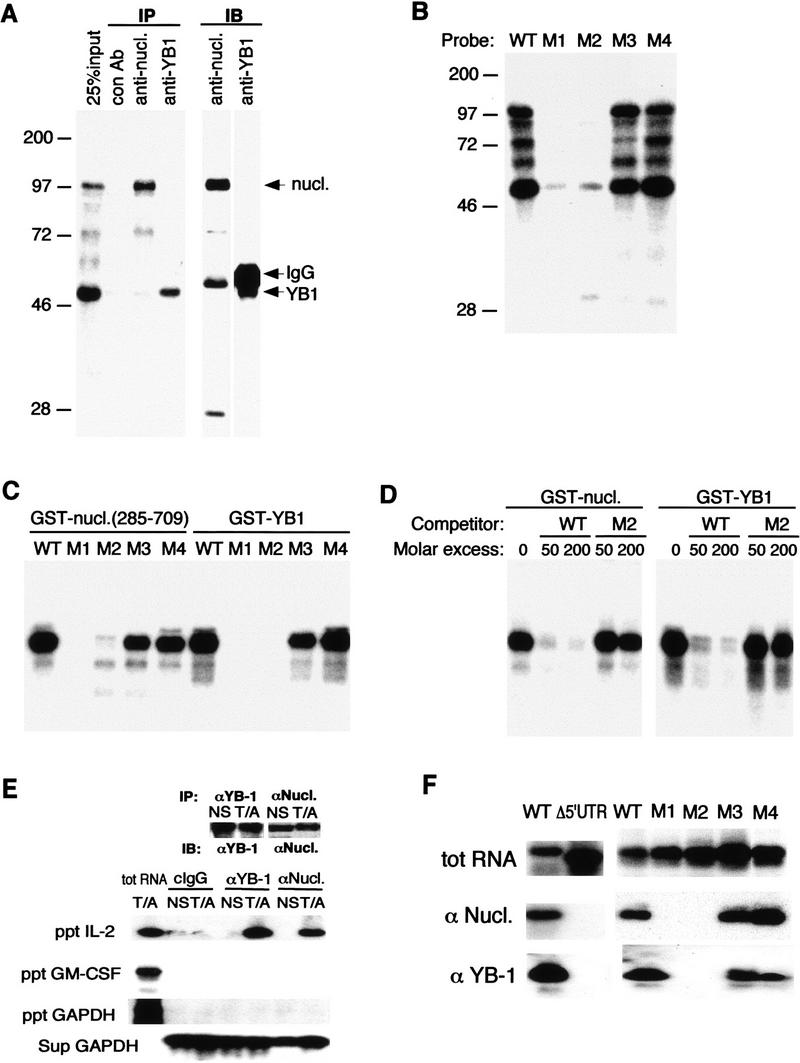
Binding of YB-1 and nucleolin to IL-2 RNA. (A) Jurkat cytoplasmic extracts were incubated with 32P-labeled IL-2(1–58) RNA, UV cross-linked, and immunoprecipitated with control antibodies [mouse IgG (lane 2) or rabbit IgG (not shown)], a monoclonal antibody against nucleolin (lane 3), or polyclonal antibodies against YB-1 (lane 4). The input (25%, lane 1) and the precipitates were fractionated by SDS-PAGE, transferred to a membrane, and autoradiographed. These membranes were then probed with anti-nucleolin (lane 5) or anti-YB-1 (lane 6) antibodies and visualized by ECL. (B) UV cross-linking of wild-type and mutant IL-2(1–58) RNAs incubated with Jurkat extracts. (C) Binding of recombinant nucleolin or YB-1 to wild-type and mutant IL-2(1–58) RNAs. Each probe was incubated with GST–nucleolin(285–709) (100 ng) or GST–YB-1 (25 ng) and analyzed by UV cross-linking. (D) Specific binding of GST–nucleolin(285–709) or GST–YB-1 to IL-2(1–58) RNA. Wild-type 32P-IL-2(1–58) RNA was incubated with nucleolin or YB-1 in the presence of molar excess of unlabeled wild-type or M2 IL-2(1–58) RNAs and analyzed by UV cross-linking. (E) In vivo association of IL-2 mRNA with nucleolin and YB-1. The proteins were immunoprecipitated from either unstimulated (NS) or stimulated (T/A) Jurkat cells. Portions of the precipitates were analyzed by immunoblotting (IB) with either anti-YB-1 or anti-nucleolin (top). RNA was extracted from the immune complexes (ppt) and analyzed by RT–PCR and Southern blotting. RNA was also extracted from supernatants (sup) and similarly analyzed. (F) In vivo association of nucleolin and YB-1 with wild-type or IL-2 mRNA mutants. Jurkat cells were transfected with vectors expressing either wild-type IL-2 mRNA or a mutant in which the IL-2 5′ UTR was removed (Δ5′-UTR) or CAT reporters, containing wild-type or mutant 5′ UTRs, described in Fig. 1D. After 48 hr, cells were stimulated, lysed, and immunoprecipitated with anti-nucleolin or anti-YB-1. The presence of reporter-derived transcripts in the immune complexes was analyzed by RT–PCR. (Top) Expression of each transcript in transfected cells; (middle, bottom) precipitated RNAs.