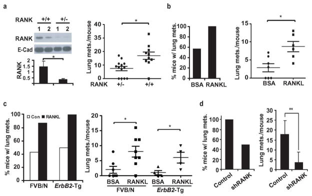
Figure 1. RANK-signaling in mammary carcinoma cells enhances metastasis.
a, MMTV-ErbB2/Rank+/+/Fvb and MMTV-ErbB2/Rank+/−/Fvb mice were sacrificed 8 weeks after tumor onset. RANK expression in primary tumors was analyzed by immunoblotting (each lane a tumor from a different mouse, upper left) and quantitated by densitometry of relative to E-cadherin (bottom left; means +/− s.e.m.; n=6). Lung metastasis multiplicity was analyzed by H&E staining of lungs from MMTV-ErbB2/Rank+/+/Fvb (n=10) and MMTV-ErbB2/Rank+/−/Fvb (n=13) mice. Means +/− s.e.m. and data for individual mice are shown (right panel). b, PCaM cells from MMTV-ErbB2/Fvb mice were isolated and grafted (1×10⁶ cells) into the #2 mammary glands of FVB/N mice subjected to twice weekly intra-tumoral injections of BSA or recombinant RANKL (80 μg/kg), starting one week post-inoculation. At day 35, lungs were isolated, sectioned, H&E stained, and metastatic lesions counted. Shown are means +/− s.e.m. (n=6) as well as values for individual mice. c, MT2 cells (1×10⁶ ) were grafted into #2 mammary glands of FVB/N or MMTV-ErbB2/Fvb mice treated with BSA or RANKL and analyzed for pulmonary metastases as in a. Data are presented as incidence and multiplicity of pulmonary metastases (mean +/− s.e.m.; n=7–8). d, Mice were inoculated with 1×10⁶ shRANK-transduced or control MT2 cells and lung metastases were measured 56 days later. Data are presented as incidence and multiplicity of pulmonary metastases (means +/− s.e.m.; n=6). *: P<0.05; **: P<0.01.

