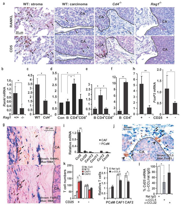
Figure 2. Expression of RANKL in mammary tumors depends on CD4+ T cells.
a, Mammary glands of indicated mouse strains were inoculated with 1×10⁶ MMTV-ErbB2 PCaM cells. After 56 days, tumors were isolated, fixed, paraffin embedded and sectioned. Parallel sections were stained with RANKL- and CD5-specific antibodies and counterstained with hematoxylin. Panels show the stroma- and carcinoma-containing regions separated by a line, when relevant. Arrows indicate CD5+ and RANKL+ cells. b–f, Rankl mRNA expression in tumors. b, MT2 cells were transplanted into the #2 mammary glands of indicated mice. After 56 days, tumors were excised and Rankl mRNA was quantitated by q-RT-PCR and normalized to cyclophilin mRNA. Results are means +/− s.e.m. (n=3). c, Freshly-isolated MMTV-ErbB2 PCaM cells were transplanted as above into indicated mice. After 56 days, tumors were isolated and Rankl mRNA was quantified. Results are means +/−s.e.m. (n=3). d, MT2 cells were transplanted into mock-, B cell-, CD4+ T cell- or CD8+ T cell-reconstituted Rag1−/− mice, 3 days after reconstitution. Rankl mRNA in tumors was analyzed as above. Means +/− s.e.m. (n=3). e, Spontaneous MMTV-ErbB2 tumors were dissociated into single cell suspensions. Tumor-infiltrating lymphocytes were enriched by positive selection and Rankl mRNA was quantified as above. Means +/− s.e.m. (n=3). f, Tumor-infiltrating B cells and CD4+ T cells were purified from MT2 formed tumors and analyzed for Rankl mRNA. Means +/− s.e.m. (n=3). *: P<0.05; **: P<0.01. g, Parallel sections of tumors raised in WT mice as in a. were stained with FoxP3 (brown)-, RANKL (brown)- and SMA (blue)-specific antibodies without counterstaining. Arrows: FoxP3+ and RANKL+ cells. The stroma and carcinoma regions are separated by a line. h, Tumor-infiltrating CD4+CD25+ and CD4+CD25− T cells were purified and analyzed for Rankl and FoxP3 mRNA expression as above. Means +/− s.e.m. (n=3). *: P<0.05; **: P<0.01. i, Cancer associated fibroblasts (CAF) and PCaM cells were purified from MMTV-ErbB2 tumors. Chemokine mRNAs were quantified by qRT-PCR as above. Means +/− s.e.m. (n=3). **: P<0.01. j, Sectioned MMTV-ErbB2 tumors were stained with CCL5- (brown) and FoxP3-(blue) specific antibodies. k, The indicated cell types were plated onto multiwell plates at 2X10⁵ cells per well with 3 μg/ml rat IgG or CCL5 antibody. Splenocytes from tumor-bearing MMTV-ErbB2 mice were added to the upper compartment of Boyden chambers. After 24 hrs, T cells in the bottom compartment were quantified by flow cytometry. CAF1 and CAF2, two independent preparations. Means +/− s.e.m. (n=3). *, P<0.05. l, Rat IgG, CCL5, or CCL22 antibodies (2 mg/kg) were i.p. injected into FVB/N females bearing MT2 tumors twice weekly. After one week, tumors were excised and Cd25 mRNA was quantified by q-RT-PCR and normalized to cyclophilin mRNA. Results are means +/− s.e.m. (n=4). *, P<0.05. CA, carcinoma region.

