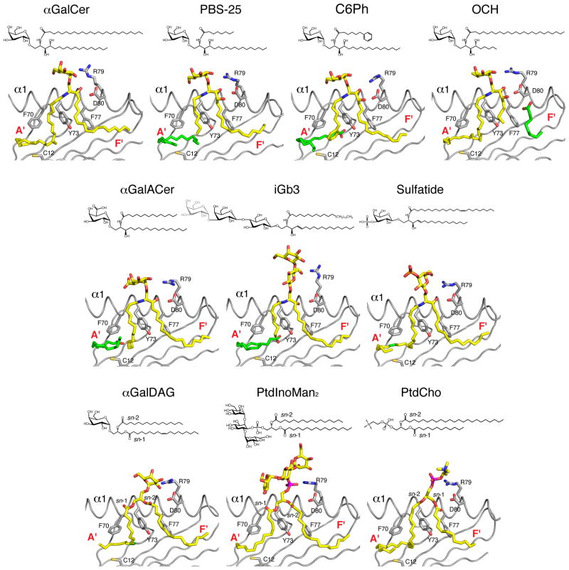
Figure 2. Chemical structure and orientation of ligands in the mCD1d ABG.
Top, chemical structures of the ligands; gray, portions not ordered in the corresponding crystal structures. Bottom, a side view of the ABG with the α2 helix removed for clarity. Ligand, yellow; spacer lipids, green; mouse CD1d heavy chain, gray; unsaturations on the acyl chains of the ligands are also green. Some of the residues involved in defining the ABG and contacting the ligand are highlighted. PDB ID: αGalCer (human), 1ZT4;αGalCer, 1Z5L; C6Ph, 3GML; OCH, 3G08; αGalACer, 2FIK; iGb3, 2Q7Y; sulfatide, 2AKR; αGalDAG, 3ILQ; PtdInoMan2, 2GAZ; PtdCho, 1ZHN.

