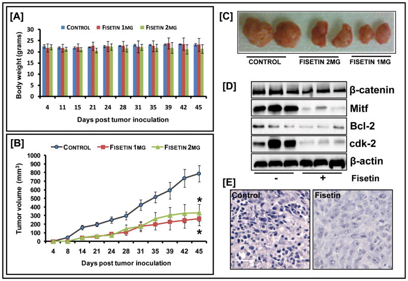
Figure 6. Effect of fisetin on 451Lu tumor growth in athymic nude mice.
(A) Average weight of control and fisetin-treated mice plotted over days after tumor cell inoculation: points; mean of 12 tumors in six animals; bars represent SD, p<0.05, versus the control group (B) Average tumor volume of control and fisetin-treated mice plotted over days after tumor cell inoculation. Points, mean of 12 tumors in six animals; bars, SEM*, p<0.001, versus the control group (C) Photographs of excised tumors from each group (D) Whole cell lysates of tumor tissues analyzed by western blot; equal loading confirmed by β-actin (E) Mitf staining in tumor sections of fisetin-treated/untreated mice (bar=25μm). Data shown is representative of samples from each group repeated thrice with similar results.

