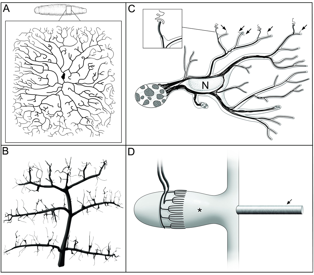
Figure 3. Multi-dendritic polymodal nociceptors and mechanosensors from different organisms.
A. Drosophila DA-IV neurons. The skin of the fly third instar larva is tiled with dendritic arbors that cover large receptive fields; the many free endings of each cell show self-avoidance. Redrawn with permission, based on immunohistochemical images from [25].
B. Leech P-cell. Pressure receptors in leech skin have distinctive multi-branched arbors with very fine endings that show self-avoidance (adapted, with permission, based on dye-filling images in [37]). Their receptive fields cover large portions of the leech skin and are known to substantially overlap circumferentially (i.e. no tiling).
C. Penicillate neuron of human skin. Nociceptors form highly branched dendrites ensheathed by a Schwann cell and its processes, except for short free endings at their termini (gray process shown in inset) that insert themselves at the borders between epidermal cells (wiggly line extending from the ending) or by invagination into a subepidermal cell. Short terminal branches of the neuron run within peripheral branches of the Schwann cell, and anchor to the epidermal basal lamina (arrows). These neurons are unmeylinated and belong to the C-fiber neuron class. N, Schwann cell nucleus. Based on schematic diagram in [30].
D. Palisade neuron of mammalian fur. Mechanosensitive dendrites end in candelabra-like arbors. These arbors show self-avoidance, and perhaps tiling, and wrap around the base of a vibrissal hair follicle (indicated by *). A single protruding hair is indicated by an arrow. Adapted, with permission, from [60].

