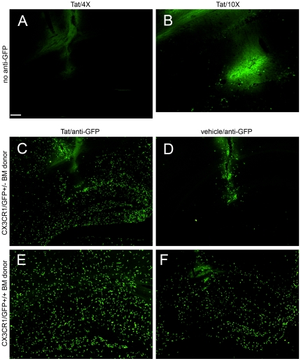
Figure 2. Stereotactic injection of Tat induces a differential effect on infiltrating inflammatory leukocytes depending on gene dose of CX3CR1.
Montage of microscopic fields 24 hours after Tat (Panels A–C, E) or control vehicle (Panels D, F) injection into the right hippocampus of irradiation chimeras. Images in Panels A, B, C, and D were taken from CD45.1 host mice engrafted with heterozygous CX3CR1/GFP+/− bone marrow, whereas images E and F were from CD45.1 host mice engrafted with homozygous CX3CR1/GFP+/+ bone marrow. A, C, D, E, and F were taken with a 4× objective with the same 0.9 sec exposure time and B was taken with a 10× objective with a longer exposure time (4 sec) to intensify the weak fluorescence of the GFP signal. Panel A and B depict very low expression of fluorescence from infiltrating GFP expressing leukocytes, while Panels C–F depict amplified GFP expression with anti-GFP antibody and Alexa 488 conjugated second antibody treatment. All experimental and control groups, n = 3 independent replicates. Scale bar = 100 µm for Panels A, C, D, E, and F; 40 µm for B.

