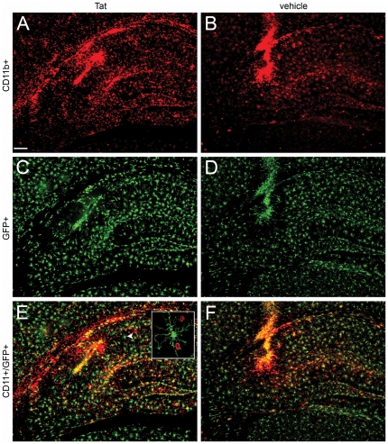
Figure 3. Stereotactic injection of Tat into hippocampus does not induce amoeboid microglial morphology in irradiation chimera.
Montage of micrographs 24 hours after Tat (Panels A, C, and E) or control vehicle (Panels B, D, and F) injection into right hippocampus in CX3CR1/GFP+/− hosts engrafted with congenic CD45.1 bone marrow. A and B show CD11b+ cells (red), C and D show GFP+ microglia (green) present in the same section, and E and F presents an overlay of the two images - highlighting the CD11b+/GFP+ co-expressing cells (orange) as well as the single positive cells. The inset in panel E shows a ramified GFP+ microglial cell (white arrowhead) in higher magnification and the 2 infiltrating CD11b+ peripheral leukocytes (red). All experimental and control groups, n = 3 independent replicates. Scale bar shown in Panel A = 100 µm for all figures.

