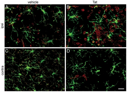
Figure 4. High resolution micrographs confirm lack of amoeboid microglial morphology following exposure to Tat in irradiation chimera.
Montage of high resolution micrographs illustrating the microglia remain in a ramified morphology 24 hours after Tat (Panels B, D) or control vehicle (Panels A, C) injection into right hippocampus in CX3CR1/GFP+/− hosts engrafted with congenic CD45.1 bone marrow. Shown are CD11b+ cells (red) and GFP+ microglia (green). While thickened processes are frequently observed, there is no evidence of amoeboid microglial morphology. Panels A and B were taken from an area of the hippocampus near the injection site of vehicle and Tat, respectively, and C and D were from the corresponding area of the hippocampus contralateral to the injection site of vehicle and Tat, respectively. All experimental and control groups, n = 3 independent replicates. Scale bar = 20 µm.

