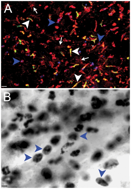
Figure 6. Stereotactic injection of Tat into hippocampus induces Infiltration of polymorphonuclear cells 24 hours later.
Panel A illustrates combined immunofluorescence staining of CD11b (red), MPO (Blue), Iba-1 (white), and GFP (green) cells in a representative tissue section from a congenic CD45.1 host engrafted with homozygous CX3CR1/GFP+/+ marrow that received Tat. Blue arrowheads point to CD11b+/MPO+/GFP− infiltrating leukocytes, white arrowheads point to CD11b+/MPO−/GFP+ infiltrating leukocytes, and white arrows point to CD11b+low/MPO−/GFP− ramified microglia. Panel B illustrates a 100× oil-immersion image from H&E staining of an adjacent section from Panel A and blue arrowheads point to polymorphonuclear cells. Scale bar = 16 and 8 µm for Panels A and B, respectively. n = 3 independent replicates for Tat treatment.

