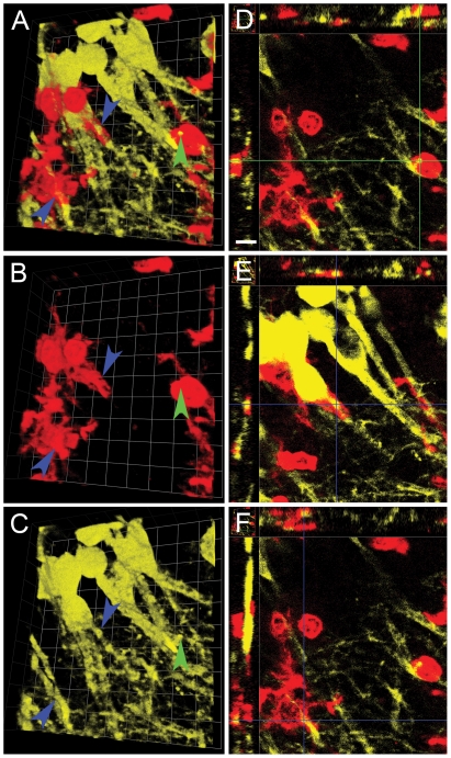
Figure 9. Stereotactic injection of Tat induces infiltrating leukocyte engulfment of dendrites.
This is a montage of 3D reconstruction micrographs of a representative tissue section of right hippocampus of YFPH mice that received Tat and were sacrificed 24 hours later (n = 3 independent replicates). As in Figure 7, Panels A–C depict a 3D reconstruction of CD11b+ microglia (red) and infiltrating leukocytes (also red) interacting with YFP+ pyramidal neurons (yellow) while Panels D–F show XYZ views at different depths in the same image stack. Green arrowhead points to an infiltrating leukocyte phagocytosing a dendritic process from an YFP+ pyramidal neuron, which is depicted in the XYZ view in Panel D (crossing point of the two green cursor lines). In contrast, blue arrowheads identify where microglia processes appear to ensheath neuronal processes. Panels E and F depict the XYZ view of this microglial ensheathment of a neuronal process (crossing point of the two blue cursor lines). Each side of individual grid = 9 µm in Panels A–C, and scale bar in Panel D = 10 µm for Panels D–F.

