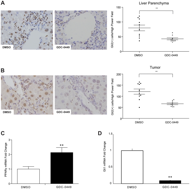
Figure 2. Hedgehog (Hh) inhibitor, GDC-0449, abrogates effects of Hh signaling within liver parenchyma and HCC nodules.
A. Liver sections stained for Gli2 from representative DMSO- and GDC-0449- treated mice (40×). Quantitative Gli2 immunohistochemistry data in non-tumor livers of mice treated with DMSO or GDC-0449 (n = 9–10/group) are graphed as mean±SEM (**p<0.01. The number of ductular cells with Gli2 positive staining were counted in each portal tract/section under 40× magnification. B. Tumor sections from the same mice were also stained to demonstrate Gli2. Results from representative DMSO- and GDC-0449-treated mice are displayed. Quantitative Gli2 immunohistochemistry data were generated by counting nuclear Gli2 positive ductular and hepatocytic cells in tumor sections under 40× magnification. Results are graphed as mean±SEM Gli2-positive cells/40× high power field (**p<0.01) C–D Quantitative reverse transcription-PCR (qRT-PCR) analysis of whole liver RNA from DMSO-(open bar) and GDC-0449 (black bar) treated mice. C. PPAR-γ, a gene that is normally repressed by Hh signaling. D. Gli1, a gene that is induced by Hh signaling. Mean±SEM are graphed (**p<0.01).

