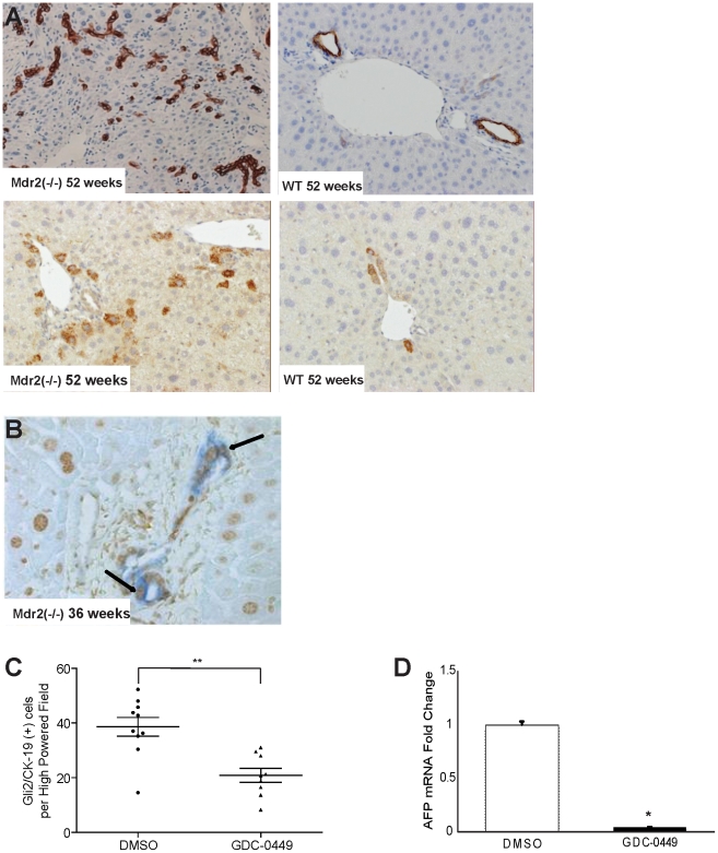
Figure 4. Effects of Mdr2 deficiency and Hedgehog (Hh) inhibition on hepatic progenitor populations.
A. Immunohistochemical staining of liver sections from representative, age-matched Mdr2−/− and wildtype mice for progenitor markers, cytokeratin-19 (CK-19) (top panels) and α -fetoprotein (AFP) (bottom panels) (20×). B. Representative micrograph of a portal triad in liver of an Mdr2−/− mouse, demonstrating co-localization of CK-19 (blue) and Gli2 (brown) in the ductular compartment (40×). C. Quantitative Gli2 and CK19 immunohistochemistry in DMSO- and GDC-0449-treated Mdr2−/− mice (n = 9–10/group). The number of Gli2 and CK19 double-positive ductular-appearing cells were counted within tumors under 20× magnification. Mean±SEM double(+) cells t per high power field (HPF) are graphed (* p<0.05) D. QRT-PCR analysis of AFP in tumor RNA from mice treated with DMSO (open bar) or GDC-0449 (closed bar) Results in the GDC-0449-treated mice were normalized to that of the mice treated with DMSO vehicle and graphed as Mean±SEM (*p<0.05).

