
Figure 10.
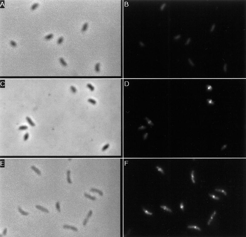
Immunolocalization of FtsZ in different cell types. Cells were synchronized and allowed to proceed through the cell cycle in M2-G medium. Samples of swarmer cells (A,B), stalked cells (C,D), and predivisional cells (E,F) were taken and processed for immunofluorescence. (A,C,E) Phase contrast micrographs; (B,D,F) the FITC immunostaining of FtsZ of the same field as shown in phase contrast micrographs.