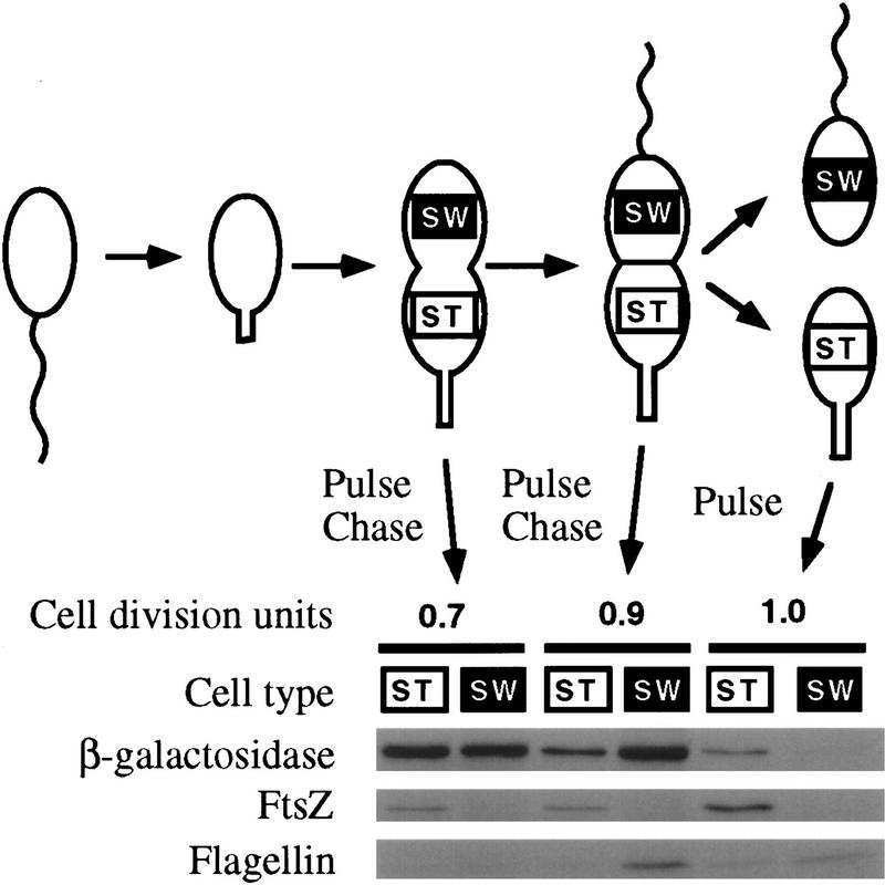
Figure 3.
Expression of FtsZ in different cell types before and after cell division. A synchronized population of swarmer cells harboring ftsZ–lacZ fusion plasmid plac290/HB2.0BP was allowed to proceed through the cell cycle. After 0.7 and 0.9 cell division unit the cells were pulse-labeled for 5 min with [35S]methionine. The label was chased with unlabeled methionine and the cells were allowed to divide. Immediately after division, swarmer (SW) and stalked (ST) cells were separated by density centrifugation. Cell extracts were immunoprecipitated with antibodies to β-galactosidase, FtsZ, and flagellins. The labeled proteins were visualized by autoradiography following SDS-PAGE. In addition, when the cells had just completed division, they were pulse-labeled for 5 min and swarmer and stalked cells were separated. The relative rates of synthesis of β-galactosidase (a measure of the rate of ftsZ transcription) and of FtsZ were measured by phosphorimaging quantitation of immunoprecipitated proteins.

