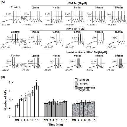
Figure 1. HIV-1 Tat enhanced excitability of small-diameter DRG neurons.
Small-diameter rat primary DRG neurons were treated with HIV-1 Tat, Gag or heat-inactivated Tat, and then subjected to whole-cell patch-clamp recordings to determine action potentials (APs). (A) A representative experiment of AP recording from one of fifteen DRG neurons is shown. The experiment was repeated at least two times. DRG neurons were exposed to 1 µM or 20 µM of Tat, 20 µM of Gag or 20 µM of heat-inactivated Tat, and the APs were recorded at 2 to 15 min of post-exposure to viral proteins. Treatment with PBS was used as a control to obtain the baseline of APs. (B) Summary data from experiments performed on all fifteen DRG neurons are shown. Bars represent SEM on the mean. Comparisons were performed between PBS and all conditions according to time points of exposure using Anova or Repeated Measures Anova. Asterisk indicates statistically significant (*p<0.05) associations between PBS and Tat treatment according to time points of exposure. CN: control with PBS treatment, APs: action potentials.

