
Figure 2.
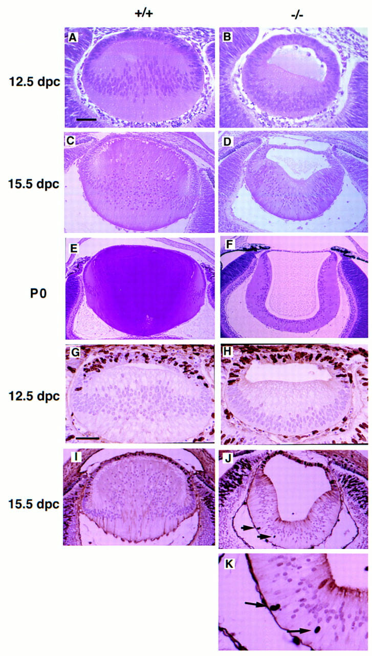
Histological analysis and cellular proliferation of wild-type and mutant lens. Hematoxylin and eosin staining of embryonic lens: (A,C,E) Wild-type lens, (B,D,F) mutant lens at 12.5 dpc (A,B), 15.5 dpc (C,D), and postnatal day zero (P0) (E,F). Loss of Sox1 causes impaired posterior lens fiber cell elongation and small, hollow lens. The nuclei in the mutant lens fiber cells are located closer to the retinal side at 12.5 dpc, whereas at P0, they are located closer to the cavity of the lens. BrdU incorporation assays: (G,I) Wild-type lens; (H,J,K) mutant lens at 12.5 dpc (G,H) and 15.5 dpc (I–K). Arrows indicate BrdU-positive nuclei in the lens fiber cells. Bar, 50 μm in A, B, G, and H; 100 μm in C and D; 140 μm in E and F. 120 μm in I and J; 50 μm in K.