
Figure 3.
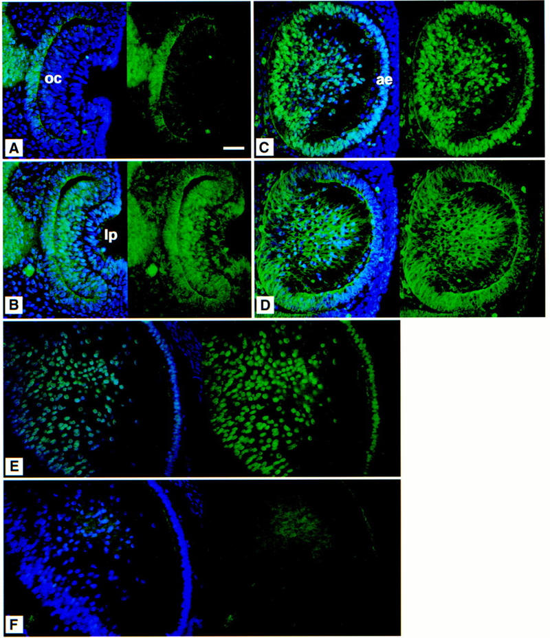
Immunofluorescent analysis of the wild-type lens. Eye sections at 10.5 (A,B), 12.5 (C,D), and 15.5 (E,F) dpc. (A,C,E) SOX1 and (B,D,F) SOX2 expression. The left of each panel shows the DAPI nuclear counterstain (blue) with SOX protein expression patterns (green); the right shows protein expression only. SOX1 protein can be detected in the nuclei of the presumptive lens fibers later during day 10 of development (not shown). SOX2 protein is detected in the nuclei of the cells of both the optic cup and the lens pit at 10.5 dpc (B). At 12.5 dpc, SOX1 is present in the nuclei of the developing lens fibers and the anterior epithelium (C); anti-SOX2 predominantly stains the cytoplasm of the lens fibers (D), although the protein is sometimes detected in the nuclei of the less differentiated lens fiber cells around the equatorial region and in the anterior epithelium (not shown). At 15.5 dpc in the lens, SOX1 is still detected in fiber cell nuclei (E), but SOX2 protein is absent (F). (oc) Optic cup; (lp) lens pit; (ae) anterior epithelium. Bar, 50 μm.