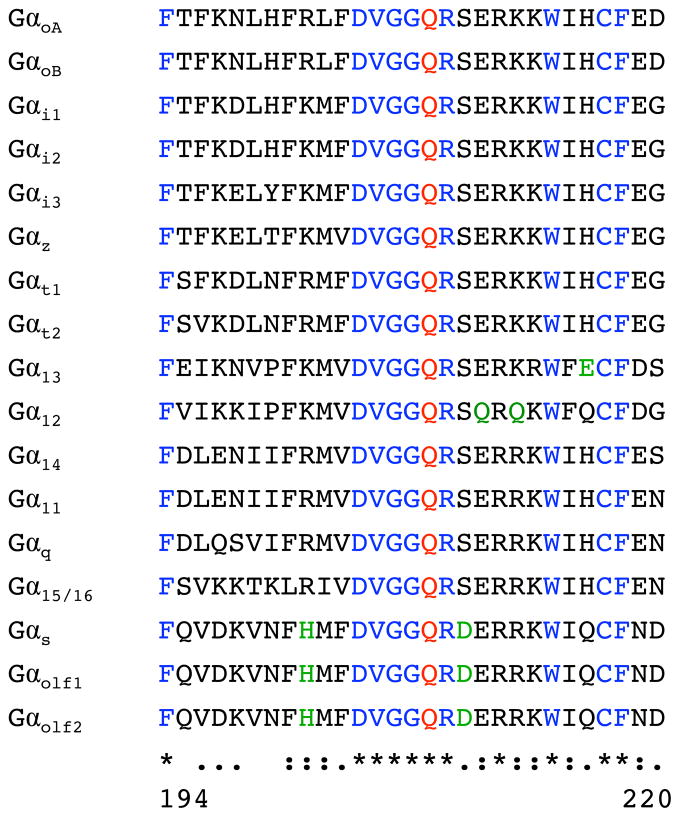
Figure 3. Alignment of amino acid sequences of the switch II region in the α subunits of heterotrimeric GTPases.
The protein sequences were obtained from NCBI; Gαt1 (NP_032166), Gαt2 (NP_032167), Gαi1 (NP_034435), Gαi2 (AAH65159), Gαi3 (NP_034436), GαoA (NP_034438), GαoB (P18873.3), Gαz (NP_034441), Gαs (P63094), Gαolf1 (NP_034437), Gαolf2 (NP_796111), Gα11 (NP_034431), Gαq (NP_032165), Gα14 (NP_032163), Gα15 (NP_034434), Gα12 (NP_034432), and Gα13 (NP_034433). The numbers below the alignment correspond to the amino acid positions of Gαq. The active site Gln (at position 209 in Gαq) is indicated in red, identical flanking residues in blue, and flanking residues that result in notable charge differences are indicated in green. “*” denotes identical amino acid residues; “:” denotes highly conserved residues; “.” denotes conserved residues.

