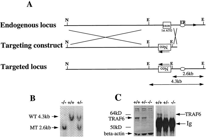
Figure 1.
Disruption of the murine traf6 gene by homologous recombination. (A). Restriction enzyme maps of the 5′ region of the endogenous traf6 locus (top) and the targeting construct (middle) are shown. The targeting vector was engineered to replace the exon containing the ATG translation initiation codon with PGK-neo in the opposite orientation (bottom). This exon encodes the first 100 amino acids of TRAF6, representing a portion of the amino-terminal ring-finger domain. EcoRI digestion generates a 4.3-kb wild-type band and a 2.6-kb mutant band. The flanking region (FR) probe used for Southern blot analysis is represented by an oval. (E) EcoRI; (N) NotI. (B) Southern blot analysis of genomic DNA from primary embryonic fibroblasts harvested from E14.5 wild-type (+/+), heterozygous (+/−) and homozygous (−/−) TRAF6 embryos. DNA was digested with EcoRI and hybridized with radiolabeled FR. Arrows indicate the 4.3-kb wild-type (WT) and the 2.6-kb mutant (MT) bands. (C) Immunoprecipitation and Western blot analysis of TRAF6 protein. TRAF6 was immunoprecipitated from total kidney lysate of TRAF6+/+, TRAF6+/−, and TRAF6−/− mice using polyclonal anti-TRAF6 antibody raised against the full-length TRAF6 protein. Western blots of both total lysates (left) and immunoprecipitates (right) were probed with the same antibody. The arrow indicates the presence of the 60-kD TRAF6 protein in +/+ lysates, a reduced level of TRAF6 in +/− lysates, and the absence of TRAF6 in −/− lysates.

