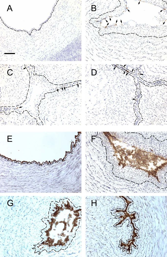
Figure 1.
VCAM-1 (A–D) and eNOS (E–H) expression in cells lining the ductus lumen. Endothelial cells were detected by eNOS expression. Ductus come from fetuses (A,E) and neonates exposed to Control conditions (B,F), anti-VLA-4 (C,G) or anti-VEGF (D,H) antibodies. Small arrows (A–D) indicate brown immunostained cells. Nuclei counterstained with hematoxylin (blue). Dashed black line = internal elastic lamina (identified by phase-contrast microscopy). Horizontal bar = 50 µm.

